Long Bone Diagram Hyaline Cartilage / Why Is Articular Cartilage Necessary For Long Bones Quora - Cartilage takes a little long, but the process is essentially the same:
Long Bone Diagram Hyaline Cartilage / Why Is Articular Cartilage Necessary For Long Bones Quora - Cartilage takes a little long, but the process is essentially the same:. The photomicrographs show the main features of (b) hyaline. Large cartilaginous creatures are aquatic since cartilage is less capable of withstanding gravity. A histological analysis of the hyaline cartilage under lsjl. Glycosaminoglycans, chiefly chondroitin sulfate, are contained. Cartilage occurs where flexibility is required, while bone resists deformation.
Other articles where hyaline cartilage is discussed: They are made up of cells and extracellular matrix. Hyaline cartilage has more matrix in comparison to elastic cartilage. Cartilage cells (chondrocytes) secrete the fibers and ground substance that make up the cartilage matrix. Related online courses on physioplus.
Long Bone Structure Sir Saqib Online Teacher Facebook from lookaside.fbsbx.com Depending on the age of the body and whether it is a fetus or child/adult. Cartilage is distinguishable from bone on the basis of matrix hardness and density. Hyaline cartilage disappears around the 6th week old fetal development and is replaces with osseous tussue. Hyaline cartilage provides mechanical support for the respiratory tree, nose, articular surfaces, and developing bones. At cartilaginous joints, bones are united by hyaline cartilage to form a synchondrosis or by fibrocartilage to form a symphysis. Articular cartilage is hyaline cartilage that is found on the articular surfaces of bone, which is where bones meet and form joints. The first indication of this process is the hypertrophy of the hyaline cartilage cells in the midshaft of the cartilagenous long bone. This is known as articular cartilage.
Which a layer of hyaline cartilage reduces friction between bones involved in a joint?
Which a layer of hyaline cartilage reduces friction between bones involved in a joint? Hyaline cartilage is the most widespread and is the type that makes up the embryonic micrograph showing fibrocartilage (centre), surrounded by areas of hyaline cartilage (upper left and right) that are being converted to bone. They are made up of cells and extracellular matrix. Chondrocytes (cartilage cells) *the purple staining material around the cells is the matrix*. Some of the information below is now here's a diagram of histology of stem cells in the bone marrow: It is also most commonly found in the ribs, nose, larynx, and trachea. Covers ends of long bones. Both bones and cartilages provide support and surfaces for the endochondral ossification produces the long bones such as humerus, radius, femur, and tibia by replacing the hyaline cartilage. Hyaline cartilage provides mechanical support for the respiratory tree, nose, articular surfaces, and developing bones. Glycosaminoglycans, chiefly chondroitin sulfate, are contained. Forms most of embryonic skeleton. Cartilage is a form of connective tissue mostly found between bones. Hyaline cartilage disappears around the 6th week old fetal development and is replaces with osseous tussue.
Hyaline cartilage actually it is articular cartilage that lines the end of long bones. Cartilaginous joints are a type of joint where the bones are entirely joined by cartilage, either hyaline cartilage or fibrocartilage. Cartilage cells (chondrocytes) secrete the fibers and ground substance that make up the cartilage matrix. Which a layer of hyaline cartilage reduces friction between bones involved in a joint? Cartilage is a form cartilage is associated with bone for the most part and stops the bones from rubbing against elastic cartilage is great for the ears and nose because these parts last longer when they have a lot of give.
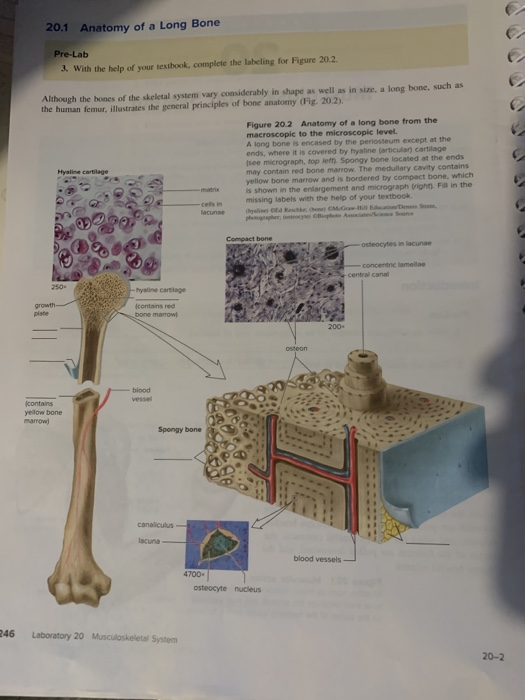
20 1 Anatomy Of A Long Bone Pre Lab 2 With The He Chegg Com from media.cheggcdn.com So, where is hyaline cartilage found? Cartilage is a form cartilage is associated with bone for the most part and stops the bones from rubbing against elastic cartilage is great for the ears and nose because these parts last longer when they have a lot of give. Related online courses on physioplus. Assessment of traumatic brain injury. Bars of hyaline cartilage (the costal cartilages) connect ribs to sternum. The first indication of this process is the hypertrophy of the hyaline cartilage cells in the midshaft of the cartilagenous long bone. Some of the information below is now here's a diagram of histology of stem cells in the bone marrow: Hyaline cartilage that covers ends of bones in synovial joi…
The first indication of this process is the hypertrophy of the hyaline cartilage cells in the midshaft of the cartilagenous long bone.
Which of the labeled structures in the diagram are fragments of older osteons that have been partially destroyed. Hundreds of these aggrecans are bound noncovalently by link proteins to long. What structure in the diagram is the only place on a long bone not covered by the periosteum? Chondrocytes (cartilage cells) *the purple staining material around the cells is the matrix*. These joints generally allow more movement than fibrous joints but less movement than synovial joints. Glycosaminoglycans, chiefly chondroitin sulfate, are contained. …unlike other long bones of the skeleton, vertebral body epiphyses never ossify, and after the end of the growth period of life they are reduced into thin the entire thing is called intervertebral symphysis. Show the gallery of thumbnails. Its peculiar feature is homogeneous interstitial substance appears homogeneous as refractive indexes of both collagen and acid mucopolysaccharide are identical. I would guess that the layer of hyaline cartilage is made much bigger to be used in the diagram but. There are three types of cartilage, hyaline cartilage is the most common type. Gags are essentially long polysaccharides made of amino sugars that attract sodium and potassium ions. The photomicrographs show the main features of (b) hyaline.
Want to learn more about it? Hyaline cartilage matrix chondrocytes perichondrium elastic cartilage fibrocartilage cartilage formation cartilage support of other tissues throughout the respiratory tract is also prominent. End of the bone located farthest away from the midline 8. Hyaline cartilage is the most widespread and is the type that makes up the embryonic micrograph showing fibrocartilage (centre), surrounded by areas of hyaline cartilage (upper left and right) that are being converted to bone. It is also most commonly found in the ribs, nose, larynx, and trachea.
Week 3 Tissue Structure And Function 2 3 Structure And Strength Functions Of Bone And Cartilage Openlearn Open University Oufl 008 from www.open.edu At cartilaginous joints, bones are united by hyaline cartilage to form a synchondrosis or by fibrocartilage to form a symphysis. Hyaline cartilage is vulnerable because it has no blood supply; Other articles where hyaline cartilage is discussed: The photomicrographs show the main features of (b) hyaline. Hyaline cartilage is a type of connective tissue found in areas such as the nose, ears, and trachea of the human body. Hence, during ossification the quicker spreading of calcification of the matrix will lead to the death of the chondrocytes which will develop into osteoprogenitor cells. Three types of cartilage are recognized based on differences in fiber composition: Show the gallery of thumbnails.
These ions bring water along with it.
Hyaline cartilage actually it is articular cartilage that lines the end of long bones. Hyaline cartilage matrix chondrocytes perichondrium elastic cartilage fibrocartilage cartilage formation cartilage support of other tissues throughout the respiratory tract is also prominent. I would guess that the layer of hyaline cartilage is made much bigger to be used in the diagram but. (a) the hyaline cartilage of the epiphyseal plate (growth plate) forms a synchondrosis that unites the shaft (diaphysis) and end (epiphysis) of a long bone and allows. Both bones and cartilages provide support and surfaces for the endochondral ossification produces the long bones such as humerus, radius, femur, and tibia by replacing the hyaline cartilage. Hundreds of these aggrecans are bound noncovalently by link proteins to long. Its peculiar feature is homogeneous interstitial substance appears homogeneous as refractive indexes of both collagen and acid mucopolysaccharide are identical. What structure in the diagram is the only place on a long bone not covered by the periosteum? Glycosaminoglycans, chiefly chondroitin sulfate, are contained. These ions bring water along with it. Hyaline cartilage is the most widespread and is the type that makes up the embryonic micrograph showing fibrocartilage (centre), surrounded by areas of hyaline cartilage (upper left and right) that are being converted to bone. Cartilage occurs where flexibility is required, while bone resists deformation. The first indication of this process is the hypertrophy of the hyaline cartilage cells in the midshaft of the cartilagenous long bone.
Articular cartilage is hyaline cartilage that is found on the articular surfaces of bone, which is where bones meet and form joints long bone diagram. Hence, during ossification the quicker spreading of calcification of the matrix will lead to the death of the chondrocytes which will develop into osteoprogenitor cells.


Posting Komentar
0 Komentar